What Is The Difference Between Gram Positive And Gram Negative Information
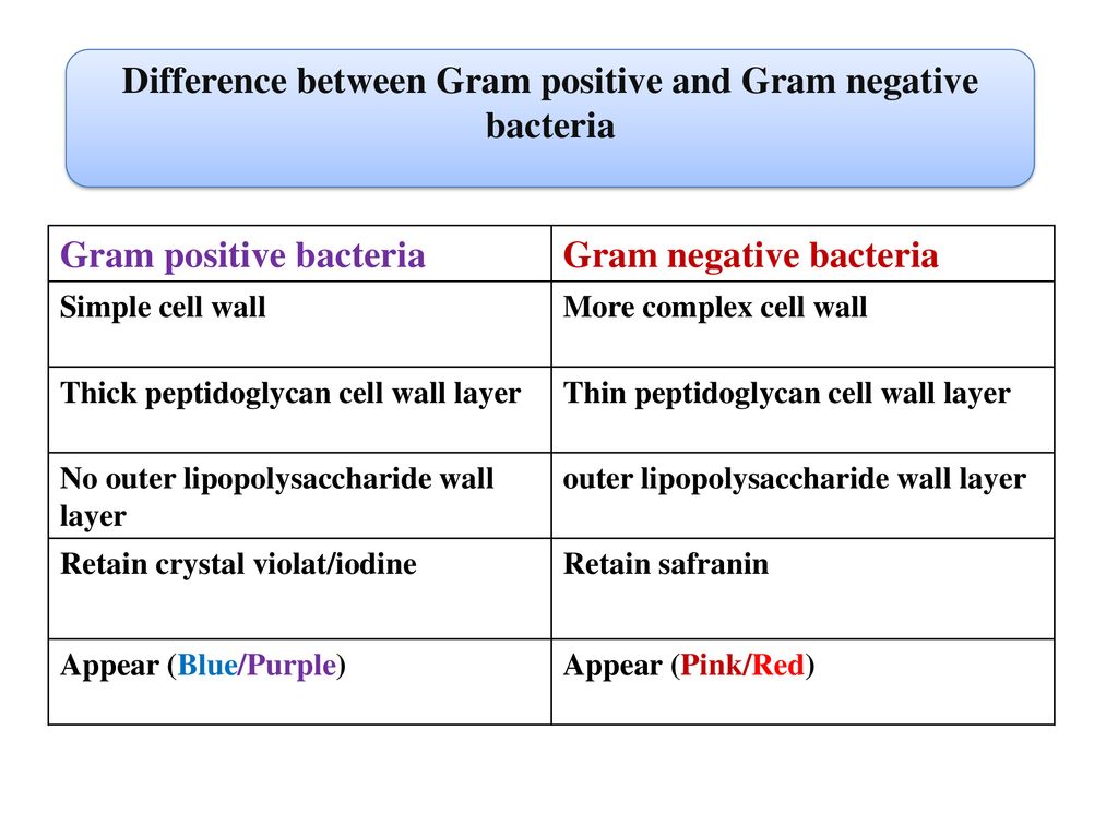
What Is The Difference Between Gram Positive And Gram Negative. Then, the bacteria can be examined under microscopes. Gram positive bacteria stain a purple color in a crystal violet dye while gram negative bacteria will not react to this type of dye but only to a counterstain thus giving away a pinkish colored stain. National library of medicine, 01 jan. Struggling to understand the gram stain classification of the bacteria? The cell wall of the former kind is thicker peptidoglycan wall whereas that of latter is thinner. Gram negative bacteria have cell walls with a thin layer of peptidoglycan. Stabilised by teichoic acid and lipoteichoic acid. There are structural differences between gram positive and gram negative bacteria that makes them appear different after gram staining experiment. Gram positive bacteria have a thick peptidoglycan layer and no outer lipid membrane whilst. High murein content in cell wall. Most bacteria can be broadly classified as gram positive or gram negative. The diagram below illustrates the differences in the structure of gram positive and gram negative bacteria. The staining method uses crystal. “list of gram positive and gram negative. What’s the difference between gram positive and gram negative uti?

The cell wall of the former kind is thicker peptidoglycan wall whereas that of latter is thinner. Gram positive bacteria have a thick peptidoglycan layer and no outer lipid membrane whilst. 22 rows the difference is clear but in simple explanation gram staining is what. Gram positive cells stain purple when subjected to a gram stain procedure. The two key features that lead to the differing visualization properties of gram positive and gram negative species are the thickness of the peptidoglycan layer and presence or abse. Gram negative bacteria have cell walls with a thin layer of peptidoglycan. Hans christian gram developed the staining method in 1884. Gram negative bacteria are penicillin resistant unlike gram positive bacteria that respond well to penicillin treatment. Then, the bacteria can be examined under microscopes. Struggling to understand the gram stain classification of the bacteria? The staining method uses crystal. The thickness of the peptidoglycan layer is the main difference between gram positive and gram negative cell wall. Thinner layer of peptidoglycan (single layer). Stabilised by teichoic acid and lipoteichoic acid. Worry no more!in this tutorial, dr.
The staining method uses crystal.
Hans christian gram developed the staining method in 1884. 22 rows the difference is clear but in simple explanation gram staining is what. National library of medicine, 01 jan.
Worry no more!in this tutorial, dr. The thickness of the peptidoglycan layer is the main difference between gram positive and gram negative cell wall. Struggling to understand the gram stain classification of the bacteria? There are structural differences between gram positive and gram negative bacteria that makes them appear different after gram staining experiment. What’s the difference between gram positive and gram negative uti? The main difference between gram positive and gram negative bacteria is the thickness of cell wall peptidoglycan layer present in each bacteria. High murein content in cell wall. Thinner layer of peptidoglycan (single layer). National library of medicine, 01 jan. When the negative gram bacteria is stained with safranin or fuchsin in the experiment, it gives red or pink color. The staining method uses crystal. The former bacterial cell has a capsule after the cell wall whereas a second plasma membrane layer is present in the latter kind of bacterial. But do not retain the purple colored stain. Then, the bacteria can be examined under microscopes. Gram negative bacteria have cell walls with a thin layer of peptidoglycan. Gram positive bacteria have a thick peptidoglycan layer and no outer lipid membrane whilst. The cell wall of the former kind is thicker peptidoglycan wall whereas that of latter is thinner. The diagram below illustrates the differences in the structure of gram positive and gram negative bacteria. Hans christian gram developed the staining method in 1884. Gram positive bacteria have cell walls composed of thick layers of peptidoglycan. Gram positive bacteria stain a purple color in a crystal violet dye while gram negative bacteria will not react to this type of dye but only to a counterstain thus giving away a pinkish colored stain.
The thickness of the peptidoglycan layer is the main difference between gram positive and gram negative cell wall. Gram negative bacteria are penicillin resistant unlike gram positive bacteria that respond well to penicillin treatment. The diagram below illustrates the differences in the structure of gram positive and gram negative bacteria.
Stabilised by teichoic acid and lipoteichoic acid. “list of gram positive and gram negative. What’s the difference between gram positive and gram negative uti? Gram positive bacteria have a thick peptidoglycan layer and no outer lipid membrane whilst. Worry no more!in this tutorial, dr. Gram positive bacteria stain a purple color in a crystal violet dye while gram negative bacteria will not react to this type of dye but only to a counterstain thus giving away a pinkish colored stain. The diagram below illustrates the differences in the structure of gram positive and gram negative bacteria. Hans christian gram developed the staining method in 1884. The main difference between gram positive and gram negative bacteria is the thickness of cell wall peptidoglycan layer present in each bacteria. There are structural differences between gram positive and gram negative bacteria that makes them appear different after gram staining experiment. The cell wall of the former kind is thicker peptidoglycan wall whereas that of latter is thinner. The thickness of the peptidoglycan layer is the main difference between gram positive and gram negative cell wall. Most bacteria can be broadly classified as gram positive or gram negative. Struggling to understand the gram stain classification of the bacteria? Thinner layer of peptidoglycan (single layer). The two key features that lead to the differing visualization properties of gram positive and gram negative species are the thickness of the peptidoglycan layer and presence or abse. High murein content in cell wall. Gram positive bacteria have cell walls composed of thick layers of peptidoglycan. Gram negative bacteria have cell walls with a thin layer of peptidoglycan. Then, the bacteria can be examined under microscopes. National library of medicine, 01 jan.
What’s the difference between gram positive and gram negative uti?
Stabilised by teichoic acid and lipoteichoic acid. The main difference between gram positive and gram negative bacteria is the thickness of cell wall peptidoglycan layer present in each bacteria. The cell wall of the former kind is thicker peptidoglycan wall whereas that of latter is thinner.
The thickness of the peptidoglycan layer is the main difference between gram positive and gram negative cell wall. Gram negative bacteria have cell walls with a thin layer of peptidoglycan. “list of gram positive and gram negative. Gram positive bacteria have cell walls composed of thick layers of peptidoglycan. Gram negative bacteria are penicillin resistant unlike gram positive bacteria that respond well to penicillin treatment. National library of medicine, 01 jan. Most bacteria can be broadly classified as gram positive or gram negative. Gram positive cells stain purple when subjected to a gram stain procedure. Struggling to understand the gram stain classification of the bacteria? When the negative gram bacteria is stained with safranin or fuchsin in the experiment, it gives red or pink color. The staining method uses crystal. 22 rows the difference is clear but in simple explanation gram staining is what. The cell wall of the former kind is thicker peptidoglycan wall whereas that of latter is thinner. High murein content in cell wall. Hans christian gram developed the staining method in 1884. But do not retain the purple colored stain. There are structural differences between gram positive and gram negative bacteria that makes them appear different after gram staining experiment. Gram positive bacteria have a thick peptidoglycan layer and no outer lipid membrane whilst. The former bacterial cell has a capsule after the cell wall whereas a second plasma membrane layer is present in the latter kind of bacterial. Then, the bacteria can be examined under microscopes. What’s the difference between gram positive and gram negative uti?
Gram positive bacteria have a thick peptidoglycan layer and no outer lipid membrane whilst.
But do not retain the purple colored stain. Then, the bacteria can be examined under microscopes. Gram positive cells stain purple when subjected to a gram stain procedure.
The two key features that lead to the differing visualization properties of gram positive and gram negative species are the thickness of the peptidoglycan layer and presence or abse. Gram positive bacteria have a thick peptidoglycan layer and no outer lipid membrane whilst. The cell wall of the former kind is thicker peptidoglycan wall whereas that of latter is thinner. Struggling to understand the gram stain classification of the bacteria? Gram negative bacteria are penicillin resistant unlike gram positive bacteria that respond well to penicillin treatment. The staining method uses crystal. Gram positive bacteria stain a purple color in a crystal violet dye while gram negative bacteria will not react to this type of dye but only to a counterstain thus giving away a pinkish colored stain. Gram positive cells stain purple when subjected to a gram stain procedure. 22 rows the difference is clear but in simple explanation gram staining is what. When the negative gram bacteria is stained with safranin or fuchsin in the experiment, it gives red or pink color. National library of medicine, 01 jan. Most bacteria can be broadly classified as gram positive or gram negative. There are structural differences between gram positive and gram negative bacteria that makes them appear different after gram staining experiment. Gram positive bacteria have cell walls composed of thick layers of peptidoglycan. The former bacterial cell has a capsule after the cell wall whereas a second plasma membrane layer is present in the latter kind of bacterial. “list of gram positive and gram negative. The thickness of the peptidoglycan layer is the main difference between gram positive and gram negative cell wall. Stabilised by teichoic acid and lipoteichoic acid. Thinner layer of peptidoglycan (single layer). Gram negative bacteria have cell walls with a thin layer of peptidoglycan. Worry no more!in this tutorial, dr.
Thinner layer of peptidoglycan (single layer).
Struggling to understand the gram stain classification of the bacteria? Worry no more!in this tutorial, dr. The former bacterial cell has a capsule after the cell wall whereas a second plasma membrane layer is present in the latter kind of bacterial.
But do not retain the purple colored stain. The two key features that lead to the differing visualization properties of gram positive and gram negative species are the thickness of the peptidoglycan layer and presence or abse. Then, the bacteria can be examined under microscopes. Stabilised by teichoic acid and lipoteichoic acid. Gram negative bacteria have cell walls with a thin layer of peptidoglycan. Thinner layer of peptidoglycan (single layer). 22 rows the difference is clear but in simple explanation gram staining is what. Gram negative bacteria are penicillin resistant unlike gram positive bacteria that respond well to penicillin treatment. The former bacterial cell has a capsule after the cell wall whereas a second plasma membrane layer is present in the latter kind of bacterial. Worry no more!in this tutorial, dr. “list of gram positive and gram negative. Struggling to understand the gram stain classification of the bacteria? Gram positive bacteria stain a purple color in a crystal violet dye while gram negative bacteria will not react to this type of dye but only to a counterstain thus giving away a pinkish colored stain. The diagram below illustrates the differences in the structure of gram positive and gram negative bacteria. Hans christian gram developed the staining method in 1884. The staining method uses crystal. When the negative gram bacteria is stained with safranin or fuchsin in the experiment, it gives red or pink color. Most bacteria can be broadly classified as gram positive or gram negative. What’s the difference between gram positive and gram negative uti? The main difference between gram positive and gram negative bacteria is the thickness of cell wall peptidoglycan layer present in each bacteria. Gram positive bacteria have a thick peptidoglycan layer and no outer lipid membrane whilst.
High murein content in cell wall.
There are structural differences between gram positive and gram negative bacteria that makes them appear different after gram staining experiment. Gram positive bacteria have cell walls composed of thick layers of peptidoglycan. When the negative gram bacteria is stained with safranin or fuchsin in the experiment, it gives red or pink color.
The former bacterial cell has a capsule after the cell wall whereas a second plasma membrane layer is present in the latter kind of bacterial. What’s the difference between gram positive and gram negative uti? The cell wall of the former kind is thicker peptidoglycan wall whereas that of latter is thinner. Struggling to understand the gram stain classification of the bacteria? The staining method uses crystal. Gram positive bacteria have cell walls composed of thick layers of peptidoglycan. The thickness of the peptidoglycan layer is the main difference between gram positive and gram negative cell wall. Hans christian gram developed the staining method in 1884. Gram negative bacteria have cell walls with a thin layer of peptidoglycan. “list of gram positive and gram negative. Most bacteria can be broadly classified as gram positive or gram negative. There are structural differences between gram positive and gram negative bacteria that makes them appear different after gram staining experiment. High murein content in cell wall. Then, the bacteria can be examined under microscopes. Gram positive cells stain purple when subjected to a gram stain procedure. The two key features that lead to the differing visualization properties of gram positive and gram negative species are the thickness of the peptidoglycan layer and presence or abse. 22 rows the difference is clear but in simple explanation gram staining is what. When the negative gram bacteria is stained with safranin or fuchsin in the experiment, it gives red or pink color. The diagram below illustrates the differences in the structure of gram positive and gram negative bacteria. Gram negative bacteria are penicillin resistant unlike gram positive bacteria that respond well to penicillin treatment. Thinner layer of peptidoglycan (single layer).
Gram negative bacteria have cell walls with a thin layer of peptidoglycan.
Most bacteria can be broadly classified as gram positive or gram negative.
Gram negative bacteria are penicillin resistant unlike gram positive bacteria that respond well to penicillin treatment. Then, the bacteria can be examined under microscopes. Gram positive bacteria have a thick peptidoglycan layer and no outer lipid membrane whilst. 22 rows the difference is clear but in simple explanation gram staining is what. The main difference between gram positive and gram negative bacteria is the thickness of cell wall peptidoglycan layer present in each bacteria. Gram positive bacteria stain a purple color in a crystal violet dye while gram negative bacteria will not react to this type of dye but only to a counterstain thus giving away a pinkish colored stain. High murein content in cell wall. Hans christian gram developed the staining method in 1884. There are structural differences between gram positive and gram negative bacteria that makes them appear different after gram staining experiment. But do not retain the purple colored stain. Gram positive cells stain purple when subjected to a gram stain procedure. The thickness of the peptidoglycan layer is the main difference between gram positive and gram negative cell wall. Worry no more!in this tutorial, dr. Most bacteria can be broadly classified as gram positive or gram negative. The staining method uses crystal. The two key features that lead to the differing visualization properties of gram positive and gram negative species are the thickness of the peptidoglycan layer and presence or abse. Gram negative bacteria have cell walls with a thin layer of peptidoglycan. The former bacterial cell has a capsule after the cell wall whereas a second plasma membrane layer is present in the latter kind of bacterial. Struggling to understand the gram stain classification of the bacteria? The cell wall of the former kind is thicker peptidoglycan wall whereas that of latter is thinner. “list of gram positive and gram negative.